
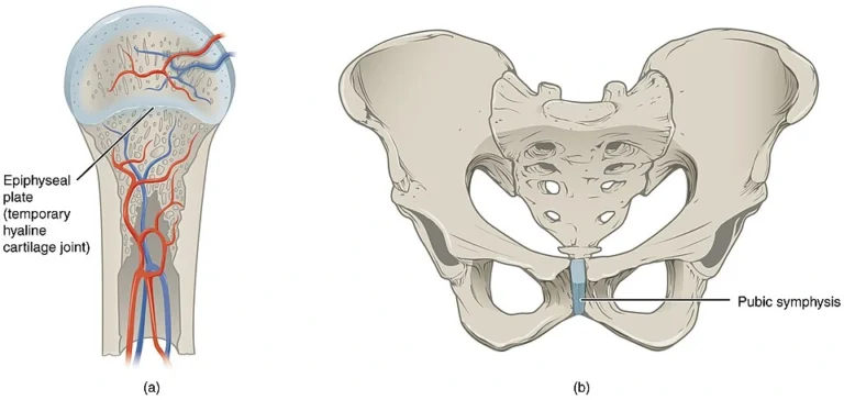
Cartilaginous Joints
Cartilaginous joints are articulations in which adjacent bones are united by cartilage rather than fibrous tissue or a synovial cavity. These joints permit more movement than fibrous joints but substantially less than synovial joints, providing a functional balance between stability and controlled flexibility. Structurally, cartilaginous joints lack a synovial cavity, and the bones are connected either by hyaline cartilage or fibrocartilage.
Cartilaginous joints are particularly important in regions where the skeleton must withstand compressive forces, absorb mechanical stress, or allow limited flexibility during growth and movement. They play a crucial role in the development of the skeleton, maintenance of axial stability, and distribution of mechanical loads within the musculoskeletal system.
Cartilaginous joints are classified into two major types based on the nature of the cartilage connecting the bones:
“Cartilaginous Joints” by OpenStax College, from Anatomy & Physiology, via Wikimedia Commons.
Licensed under CC BY 3.0
Definition
Synchondroses are cartilaginous joints in which bones are united by hyaline cartilage.
These joints are typically temporary structures that function as growth centers during skeletal development.
Structure
In a synchondrosis:
The articulating bone surfaces are connected by a layer of hyaline cartilage
The cartilage may later ossify and convert into bone
The joint therefore often becomes a synostosis (bony fusion) in adulthood
Because of the rigidity of hyaline cartilage, synchondroses generally allow little or no movement, serving primarily as structural growth zones rather than functional articulations.
Types
Epiphyseal Growth Plates (Epiphyseal Cartilage)
The most important example of a synchondrosis.
Located between the epiphysis and metaphysis of long bones
Responsible for longitudinal bone growth during childhood and adolescence
Eventually replaced by bone during epiphyseal closure
First Sternocostal Joint
Connection between the first rib and the manubrium of the sternum
Provides strong attachment with minimal movement
Spheno-occipital Synchondrosis
Located at the base of the skull
Important growth site during cranial development
Fuses during adolescence
Functional /Clinical
Functional
Synchondroses are essential for skeletal growth and developmental remodeling. They permit bones to increase in length while maintaining structural continuity between skeletal elements.
Their principal roles include:
Enabling longitudinal growth of long bones
Providing temporary structural connections during development
Allowing controlled expansion of the skeleton during childhood
Clinical
Growth Plate Injuries
Trauma affecting the epiphyseal plate can disrupt normal endochondral ossification, leading to:
limb length discrepancies
angular deformities
premature closure of the growth plate These injuries are classified clinically using the Salter–Harris classification.
Definition
Symphyses are joints in which bones are connected by fibrocartilage, usually with thin layers of hyaline cartilage covering the articular surfaces of the bones.
Unlike synchondroses, symphyses are permanent joints that persist throughout life and provide both strength and limited mobility.
Structure
Structural Characteristics
A typical symphysis consists of:
Hyaline cartilage covering the articular bone surfaces
A central fibrocartilaginous disc or pad connecting the bones
Strong surrounding ligaments reinforcing the joint
Fibrocartilage contains dense collagen bundles, enabling it to resist both compressive and tensile forces.
Types
Intervertebral Joints (Intervertebral Discs)
Located between the vertebral bodies of the spine.
Each disc consists of:
Annulus fibrosus – outer fibrocartilaginous ring
Nucleus pulposus – central gelatinous core
functions include:
allowing flexibility of the vertebral column
absorbing compressive forces
distributing loads during movement
Pubic Symphysis
A fibrocartilaginous joint connecting the left and right pubic bones.
Characteristics:
reinforced by strong ligaments
capable of limited movement
slightly increases mobility during childbirth
Functional /Clinical
Functional
Symphyses perform several important mechanical functions:
Allow slight but controlled movement between bones
Absorb shock and compressive forces
Maintain stability of the axial skeleton
Distribute mechanical loads during movement
Because fibrocartilage is highly resistant to stress, symphyses are typically located in weight-bearing or load-transmitting regions.
Clinical
Intervertebral Disc Degeneration
Degeneration or herniation of intervertebral discs can compress spinal nerves, producing conditions such as:
lumbar disc herniation
radiculopathy
sciatica
Pubic Symphysis Dysfunction
Excessive mobility or inflammation of the pubic symphysis may occur during pregnancy or trauma, causing pelvic instability and pain.
Bone Growth
Cartilaginous joints—particularly synchondrosesv – serve as dynamic growth interfaces that enable longitudinal bone development.
The epiphyseal growth plate provides a highly regulated zone of chondrocyte proliferation, hypertrophy, and matrix mineralization, facilitating endochondral ossification and determining final bone length and morphology.
Shock Absorption
Cartilaginous joints act as biomechanical buffers, especially within the axial skeleton. Fibrocartilaginous structures (e.g., intervertebral discs) are specialized to resist compressive, tensile, and shear forces, distributing loads across broader surfaces and minimizing peak stress. This protects osseous structures from mechanical fatigue and microdamage.
Controlled Flexibillity
These joints provide a functional balance between rigidity and mobility, allowing limited but essential movement while preserving skeletal continuity.
This controlled compliance prevents excessive rigidity seen in fibrous joints and excessive mobility seen in synovial joints, ensuring stability without loss of adaptability in weight-bearing regions.
Force Integration
Cartilaginous joints function as integrative interfaces, linking skeletal segments into continuous mechanical units.
They enable coordinated transmission of forces across regions (e.g., vertebral column, pelvic ring), ensuring harmonized biomechanical behavior during locomotion, posture maintenance, and load-bearing activities.
Growth Disorders
Damage to synchondroses, particularly the epiphyseal plates, disrupts normal endochondral ossification, leading to growth arrest, limb length discrepancies, or angular deformities.
These injuries are clinically critical in pediatric populations due to their long-term impact on skeletal development.
Disk Degeneration
Degeneration of fibrocartilaginous symphyses (e.g., intervertebral discs) leads to loss of disc height, reduced hydration, and structural weakening, resulting in conditions such as disc herniation and spinal instability.
This impairs load distribution and may compress neural structures, causing pain and neurological deficits.
Pelvic Instability
Alterations in cartilaginous joints such as the pubic symphysis can result in pelvic instability, particularly during pregnancy or trauma.
Excessive mobility or degeneration compromises force transmission across the pelvic ring, affecting gait mechanics and weight-bearing stability.
Load Failure
Compromise of cartilaginous joints disrupts their buffering capacity, leading to abnormal stress concentration on adjacent bones and joints. This accelerates degenerative processes, contributes to overuse injuries, and reduces overall biomechanical efficiency of the musculoskeletal system.