
Joints Classification
Joint classification in arthrology establishes a systematic framework for organizing articulations according to their connective tissue composition and biomechanical behavior. Joints function as specialized interfaces between bones, integrating the skeleton into a unified mechanical system that permits load transmission, controlled mobility, and structural stability.
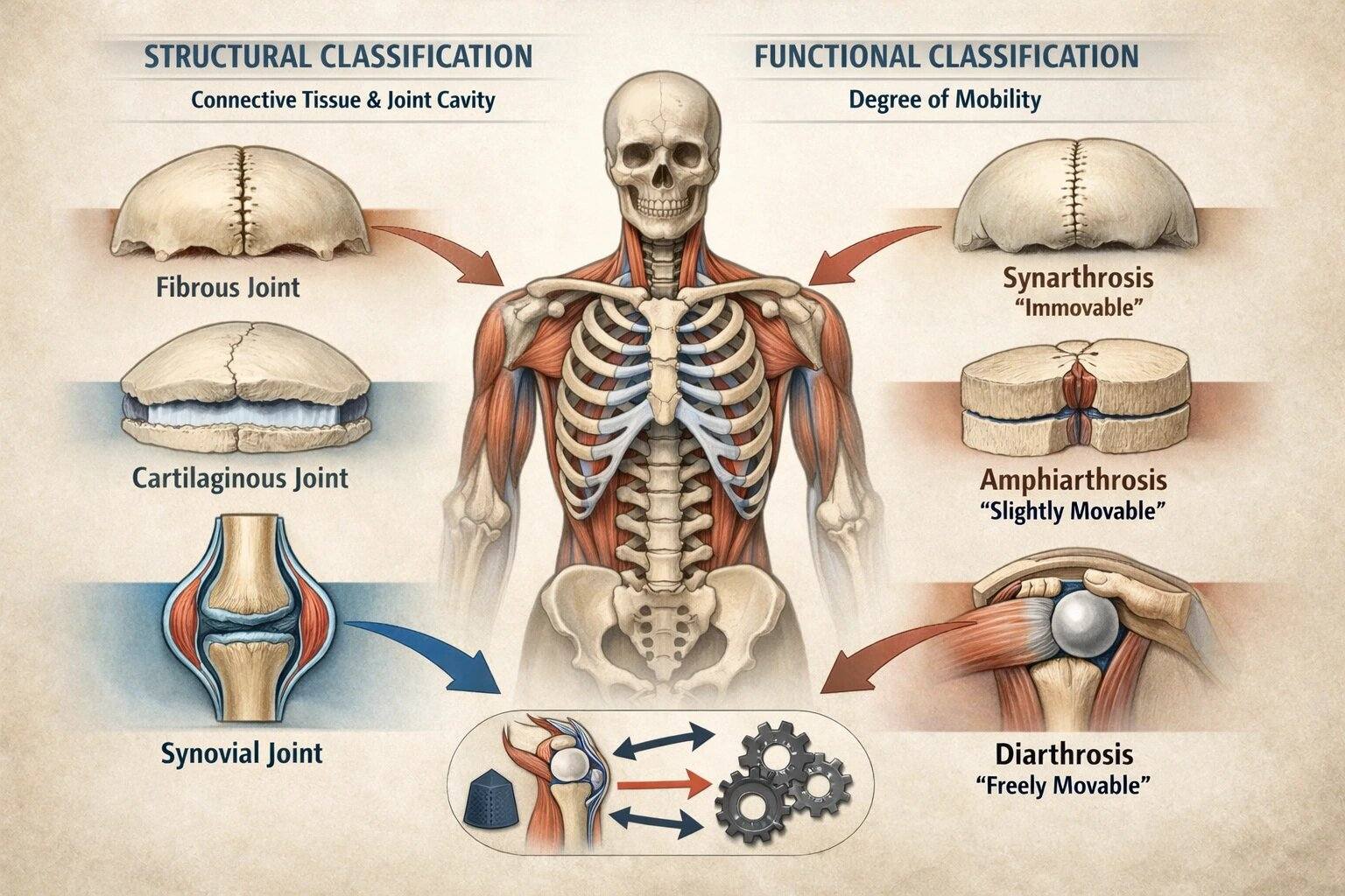
Two complementary systems are recognized. Structural classification is defined by the type of tissue uniting the bones and the presence or absence of a synovial cavity, categorizing joints into fibrous, cartilaginous, and synovial types. Functional classification is based on the degree of mobility permitted between articulating surfaces, ranging from synarthrosis (immobile) through amphiarthrosis (limited movement) to diarthrosis (freely mobile).
These systems are functionally inseparable, as structural design determines mechanical performance. The material properties of connective tissue, the geometry of articular surfaces, and the presence of a synovial cavity collectively regulate degrees of freedom, load distribution, and joint kinematics. Thus, joint classification is not purely descriptive but predictive, allowing inference of movement capacity, stability, and clinical behavior from anatomical structure.
AI-generated illustration (MyoAnatomy)
STRUCTURAL CLASSIFICATION
AI -generated illustration (MyoAantomy)
Overveiw
Joint classification provides a fundamental framework for understanding the biomechanical behavior of the musculoskeletal system. By defining joint structure, it determines the degrees of freedom available at each articulation, including the axes and range of motion through which movement occurs. It also governs the distribution and transmission of mechanical loads, ensuring that forces generated by muscular activity are efficiently transferred, absorbed, and dissipated across skeletal structures.
Furthermore, joint classification reflects the functional balance between mobility and stability. Highly mobile joints permit extensive movement at the expense of stability, whereas structurally rigid joints prioritize mechanical resistance and load-bearing capacity. This classification also underpins the integration of joints within kinematic chains, enabling coordinated, multiaxial movement through interaction with surrounding musculature.
Thus, joints function as dynamic mechanical units in which structural design dictates functional capacity, integrating anatomical constraints with biomechanical demands under physiological conditions.
Exam Question
In a biomechanical analysis of the musculoskeletal system, how does joint classification determine the relationship between structural design, degrees of freedom, and load transmission, and how do these factors collectively influence the balance between mobility and stability within kinematic chains?
Fibrous
Fibrous joints consist of bones united by dense fibrous connective tissue without the presence of a synovial cavity. These articulations exhibit minimal to no movement, with their primary function being mechanical stability, force transmission, and protection of adjacent structures. The rigidity of fibrous joints reflects their role in resisting tensile forces while maintaining structural integrity.
Three subtypes are recognized. Sutures are immobile joints between cranial bones composed of short collagen fibers; they permit slight compliance during development and contribute to protection of the brain. Syndesmoses involve bones connected by ligaments or interosseous membranes, allowing limited movement while maintaining stability and facilitating force distribution across adjacent bones. Gomphoses are specialized peg-in-socket articulations between teeth and alveolar bone, stabilized by the periodontal ligament, permitting minimal movement to absorb and dissipate mechanical stress during mastication.
Exam Question
In a biomechanical context, how does the dense fibrous connective tissue architecture of fibrous joints determine their limited mobility, and how does this structural design contribute to force transmission, tensile resistance, and protection of vital structures?
Cartilaginous
Cartilaginous joints unite bones through cartilage, providing a balance between stability and limited mobility. These joints are specialized to withstand compressive forces, absorb mechanical stress, and permit controlled flexibility without the presence of a synovial cavity.
Two subtypes are distinguished. Synchondroses are joints composed of hyaline cartilage, often temporary and associated with skeletal growth, such as the epiphyseal plates; they facilitate longitudinal bone growth and later ossify to form synostoses. Symphyses consist of fibrocartilage and are characterized by high tensile strength and shock-absorbing capacity, as seen in intervertebral discs and the pubic symphysis; they permit slight movement while effectively resisting compression and shear forces.
Exam Question
How does the composition of cartilage (hyaline vs fibrocartilage) in cartilaginous joints determine their ability to resist compressive and shear forces, and how does this structural specialization support both skeletal growth and controlled mechanical flexibility?
Synovial
Synovial joints are characterized by the presence of a fluid-filled synovial cavity separating the articulating bones, enclosed within a fibrous capsule. This structural organization permits free movement (diarthrosis) and defines synovial joints as the principal sites of dynamic, multiaxial motion within the musculoskeletal system.
Their function is governed by specialized structural components. Articular (hyaline) cartilage provides a smooth, low-friction surface that facilitates movement and distributes compressive loads across joint surfaces. The synovial membrane produces synovial fluid, which reduces friction, nourishes avascular cartilage, and enhances movement efficiency through lubrication. The fibrous capsule and associated ligaments confer mechanical stability while constraining and guiding motion within defined limits. Periarticular structures, including tendons, bursae, and fibrocartilaginous elements such as menisci or labra, further optimize joint function by improving load distribution, reducing friction, and enhancing stability.
Synovial joints are additionally classified according to the shape of articulating surfaces and the axes of movement, including hinge, pivot, condyloid, saddle, and ball-and-socket types. These configurations determine the degrees of freedom, reflecting the joint’s specialized biomechanical role in coordinating movement within kinematic chains.
Exam Question
How do the structural components of synovial joints – particularly articular cartilage, synovial fluid, and capsuloligamentous structures – interact to regulate friction reduction, load distribution, and degrees of freedom, and how does this integration enable efficient multiaxial movement while maintaining joint stability?
FUNCTIONAL CLASSIFICATION
AI -generated illustration (MyoAantomy)
Overveiw
Functional classification categorizes joints according to the degree of movement permitted between articulating bones, providing a direct reflection of their role in balancing mobility and stability within the musculoskeletal system. This system emphasizes the functional consequences of structural design, linking anatomical organization to biomechanical performance.
Joints are classified into synarthrosis (immobile), amphiarthrosis (slightly mobile), and diarthrosis (freely mobile). These categories represent a continuum in which increasing mobility is associated with decreasing intrinsic stability, requiring greater reliance on dynamic stabilizing mechanisms, including muscles and ligaments.
From a biomechanical perspective, functional classification determines the degrees of freedom available at a joint, the range and axes of motion, and the capacity for load transmission and absorption. Immobile joints prioritize structural integrity and protection, whereas highly mobile joints facilitate complex, multiaxial movement at the cost of reduced passive stability.
Thus, functional classification provides a predictive framework in which the extent of permitted motion directly reflects the mechanical demands placed on the joint, integrating structural constraints with dynamic functional requirements within kinematic chains.
Exam Question
How does functional classification of joints reflect the relationship between degree of mobility, mechanical stability, and degrees of freedom, and how do these factors influence the distribution of forces and coordination of movement within musculoskeletal kinematic chains?
Synarthrosis
Synarthrotic joints are functionally immobile articulations specialized for maximum structural stability and protection. They are most commonly associated with fibrous joints, in which dense connective tissue rigidly unites adjacent bones, eliminating movement and resisting mechanical deformation.
Biomechanically, synarthroses are optimized to withstand tensile and compressive forces without displacement, thereby maintaining structural integrity under load. Their rigidity ensures effective force transmission across skeletal units while protecting critical structures.
Representative examples include cranial sutures, where interdigitating fibrous joints stabilize the skull and protect the brain, and gomphoses (tooth–alveolar joints), where the periodontal ligament permits minimal micro-movement to absorb occlusal forces during mastication without compromising overall stability.
Thus, synarthrotic joints represent the extreme of the stability – mobility continuum, prioritizing structural cohesion over movement.
Exam Question
In the context of cranial sutures and gomphoses, how does the fibrous connective tissue architecture of synarthrotic joints enable force transmission and resistance to mechanical stress, while allowing minimal functional compliance in specific regions such as the periodontal ligament?
Amphiarthrosis
Amphiarthrotic joints permit limited movement and are structurally adapted to balance stability with controlled flexibility. They are typically associated with cartilaginous joints, particularly those composed of fibrocartilage, which allows elastic deformation under mechanical load.
Biomechanically, these joints are specialized to absorb compressive forces, redistribute mechanical stress, and maintain continuity between skeletal elements. The presence of fibrocartilage enables viscoelastic behavior, allowing the joint to deform slightly under load and return to its original shape, thereby facilitating shock absorption and load distribution.
Representative examples include the intervertebral discs, where fibrocartilage permits controlled deformation during axial loading to protect the spinal column, and the pubic symphysis, which allows limited flexibility while maintaining pelvic stability under weight-bearing conditions.
Thus, amphiarthroses occupy an intermediate position within the stability – mobility continuum, integrating structural support with limited motion to optimize mechanical efficiency in load-bearing regions.
Exam Question
In the context of intervertebral discs and the pubic symphysis, how does the fibrocartilaginous composition of amphiarthrotic joints enable viscoelastic deformation under compressive load, and how does this property contribute to shock absorption, load distribution, and controlled mobility in weight-bearing structures?
Diarthrosis
Diarthrotic joints are freely movable articulations corresponding to synovial joints, and represent the primary sites of dynamic, multiaxial movement within the musculoskeletal system. Their structural organization enables a high degree of mobility while maintaining functional stability through coordinated passive and active mechanisms.
Biomechanically, diarthroses are characterized by the presence of a synovial cavity, which permits low-friction movement, and by specialized structures that regulate motion. These joints allow a wide range of movement defined by their degrees of freedom and axes of rotation, enabling complex kinematic patterns such as flexion–extension, rotation, and circumduction. Mobility is facilitated by articular cartilage and synovial fluid, which reduce friction and distribute loads, while stability is maintained through capsuloligamentous structures and dynamic muscular control.
Representative examples include the shoulder joint, which permits extensive multiaxial movement at the expense of intrinsic stability, the hip joint, which balances mobility with structural stability for weight-bearing, and the knee joint, which enables controlled movement while managing high mechanical loads during locomotion.
Thus, diarthrotic joints represent the extreme of the mobility–stability continuum, where increased movement capacity necessitates greater reliance on dynamic stabilizing systems.
Exam Question
In the context of the shoulder, hip, and knee joints, how does the synovial joint architecture enable multiaxial movement through defined degrees of freedom, and how do articular structures and dynamic muscular stabilization interact to maintain joint stability under varying mechanical loads?
FUNCTIONAL ROLE
Motion Capacity
Joint classification defines the degrees of freedom (DOF) available at each articulation, determined by joint structure and geometry. This establishes the axes and planes of motion, constraining or permitting movement patterns from immobility to multiaxial motion. Consequently, joint type directly dictates the kinematic potential of a region within the musculoskeletal system.
Force Distribution
Joint structure governs how mechanical forces are transmitted, distributed, and dissipated across skeletal elements. Rigid joints channel forces efficiently with minimal deformation, whereas more compliant joints allow controlled deformation, enabling absorption and redistribution of stress. This ensures mechanical continuity while preventing localized overload.
Stability vs Mobility
Joint classification reflects a continuum between structural stability and functional mobility. Highly stable joints prioritize resistance to mechanical stress and preservation of integrity, while mobile joints permit greater movement at the expense of intrinsic stability. This balance enables the system to meet both support and movement demands under physiological conditions
Movement Integration
Joints function as coordinated units within integrated kinetic chains, where movement at one articulation influences adjacent segments. Classification determines how joints contribute to sequential, multiaxial motion, enabling efficient force transfer between muscles and skeletal elements. This integration underlies smooth, controlled, and purposeful movement.
CLINICAL RELEVANCE
Diagnostic Framework
Joint classification provides the structural basis for diagnosing musculoskeletal disorders, as each joint type exhibits characteristic patterns of pathology. Understanding joint architecture allows precise identification of degenerative, inflammatory, and mechanical abnormalities.
Imaging Interpretation
Knowledge of joint classification is essential for interpreting radiological findings, including joint space, alignment, and surface integrity. Structural differences between joint types determine how pathology appears across imaging modalities.
Treatment Strategy
Joint type directly influences therapeutic decision-making, including conservative management, surgical approach, and fixation techniques. Structural and biomechanical properties determine healing capacity and intervention strategy.
Injury Patterns
Different joints exhibit predictable injury patterns based on their biomechanical properties. Highly mobile joints are prone to instability, while rigid joints are more susceptible to structural failure under excessive load.